As I'm sure you can tell from the title, this was one of the most action-packed days in lab so far. In addition to running two separate projects today, it was also Jalen's birthday, the fourth one since I arrived here in June.
I got right to work when I got in around 8:15, since I had to run a glucose uptake trial. This trial was my first using the glucose strips and meter which I borrowed from Dr. Johannes' lab yesterday. Today's trial used untransfected cells because I wanted to ensure that the glucose strip protocol worked for me before using the expensive plasmids. I placed a very small amount of media on each glucose strip at intervals of one hour, and saw that there was indeed a decrease in glucose concentration of the media, indicating that the cells were taking up glucose as predicted.
In addition to the glucose uptake trial, I also ran a Western blot today. I was looking for levels of glucokinase, an enzyme that is involved in the early steps of glycolysis and helps convert glucose to energy. Therefore, this enzyme is related to the glucose uptake experiment (my original). I transfected cells with the Hepatitis B Virus (HBV) and the *7 plasmid (HBV genome without the HBx protein). My lab focuses on HBV, specifically HBx, so these two plasmids are widely used. If the HBV plasmid changes the levels of a protein while the *7 plasmid has no change, we can infer that HBx is responsible for this change.
In the middle of the glucose uptake trial and while the gels were running, we decided to eat a five-star lunch at Chik-Fil-A for Jalen's birthday. It's a fairly short walk from the lab to the mall where Chik-Fil-A is located, and we made it back in time to check on our experiments.
Tomorrow is the last day for both Jalen and Sidney. Their program will be in Philadelphia until next Friday, but they have to present early next week, and they aren't required to come back into lab after their presentation. We've had a pretty fun time in lab together, so tomorrow is a bit of a bittersweet moment. Since they are both from Texas, we probably won't see each other much after tomorrow, but we made sure to follow each other on Instagram, so I'm sure we'll keep in touch!
Thursday, July 30, 2015
Jay Ha #3 advanced oxidation
I spent the past ten days or so doing number of experiments related to advanced oxidation of OH free radicals. Purpose of these experiments was to understand the kinetics and mechanism of advanced oxidation process that I would be using next week when I am conducting UV disinfection of E. Coli using hydrogen peroxide oxidation processes.


Four experiments were conducted this past week. First one was to find experimental concentration of hydrogen peroxide. Although the concentration of h2o2 can be easily calculated using the equation of 10*d*%/molecular weight, hydrogen peroxide tends to decompose itself after the container is initially unsealed, so it was necessary to find the concentration through experiments. The calculated value was 9.8M while the experimental average was 10.486M so this value was used for further experiments. 


Second set of experiments were the decomposition hydrogen peroxide. Hydrogen peroxide were put to these  equipment containing 6, 4w UVC lamps. I extracted samples at varying time periods to see the rate at which h2o2 is decomposing since as 1 mole of h2o2 decomposes 2 moles of OH radicals are being created. This experiment was done in three varying concentrations of H2O2 solutions and the experiment was conducted twice to evaluate the error bounds.
equipment containing 6, 4w UVC lamps. I extracted samples at varying time periods to see the rate at which h2o2 is decomposing since as 1 mole of h2o2 decomposes 2 moles of OH radicals are being created. This experiment was done in three varying concentrations of H2O2 solutions and the experiment was conducted twice to evaluate the error bounds.
Third experiment dealt with the production of 4-HBA acid. This was conducted because as produced OH radicals from decomposition of H2O2 combines with benzoic acid to produce 4-HBA and this value enables researcher to estimate the amount of OH radicals that are being produced and reacting to form other compounds.
Last experiment was the decomposition of methleyne blue dye, and this was conducted because I can visually see the waste material (methleyen blue) being inactivated by OH radicals that decomposed from H2O2.
This week was more tiring than other weeks in that there were so many data and graphs to work on, but it was kind of fun as well.
Monday, July 27, 2015
David, Entry #5, Fourth of July Madness
While I was very excited for the 4th since my birthday happens to be on the 3rd, everyone I work with dreads the day. The Fourth is the one day that all humanity at the shore seems to go crazy in a "independence 'Merica" kind of way that leads to people getting drunk and doing stupid stuff on beaches. If they did this in a vacuum all would be fine, I guess, but they don't, so all the stupid things people do during the holiday haze negatively impacts plovers. Each biologist I work with gets assigned a segment of beach to patrol to call the police if anyone is disturbing the plovers. While everyone else in this country is celebrating, these very dedicated biologists are out late into the night protecting birds. I commend them for their dedication. At Belmar a group of guys decided to set a box of firecrackers off mere feet from a colony of terns. At North Brigantine a guy decided to drive around the vehicle barrier and do donuts on the beach, tearing up and destroying sensitive habitat. We also lost chick the day he illegally drove on the beach, so we highly suspect he ran it over. All across the state biologists reported horror stories like this of arrogant people harming wildlife in the name of "freedom." I'll quote spider man by saying, "with great power comes great responsibility." Sadly, the vast majority of people living at the Jersey shore seem to ignore the responsibility part. They destroy the environment they live in and then wonder why their house is flooded during storms. Our earth is deeply connected. You cannot change one thing without changing many many others. In response to the person who drove around the vehicle barrier at North Brigantine, we moved the vehicle barrier a quarter mile further down on the beach. Many town residents drove up to us as we were doing this and very vocally expressed their anger. Dealing with the general public is a very stressful and frustrating part of my work, but it's very important.


Pictured above is the vehicle barrier we built. This took about 2 hours of hard work. The barrier is created by putting large cedar posts in the sand in intervals smaller than the width of a car. Yellow cord with orange flags is strung across. Signs are put on the posts that say "endangered species area, keep out!"
As I've mentioned in a previous post, studying endangered species has its ups and downs. There is a strong emotional component that has slowly been developing in me. Last week it really seemed to hit me. We were re-capturing chicks at Holgate. We like to give all of our plovers names that we use in the field. The parents of this particular nest are Ross and Paula. The two chicks were Tuna and Noodle. However, when we got to the nest site Emily said something along the lines of, "we need to catch Ross' chick." I really didn't think anything of it until I had Tuna in my hand and realized Noodle was missing. I looked in the cat carrier we put the plovers in while we process them. Empty. Noodle had been taken only a day or two prior by a predator, yet the week before I held Noodle in my hands. I looked down into my palms as if Noodle would suddenly appear, unharmed. I could hear the trill coming from her in my memory. I could see her squirming in between my fingers. Well, I guess that's the whole point of my work: to better protect plovers in the future. If anything, it served as motivation. We work in some pretty bad conditions. There are thousands of insects that constantly bite us all day everyday. We've been caught in a few thunderstorms on the open beach with metal equipment, very very scary, especially because a lighting bolt hit the ground about 20 feet away from us the other day. One thing I've noticed about the people I work with is their extreme devotion to these birds that allows them to work through these tough circumstances. I too am developing such motivation.
Since I've talked about two sad topics on this post thus far, I'm going to end on a happy note. Enjoy the following pictures of adorable plover chicks!

 |
| Meet the Harry Potter nest! Their names are Serius, Severis, Hedwig, and Dobby (not pictured). We didn't realize until after the fact that we named them all after characters that die...Hopefully there will be some reverse psychology with the predators and they will all live! |
 |
| Chicks snuggling with their dad may be the cutest thing I've ever seen! Notice the nano tag on the chick to the far right. That allows us to track the birds 24/7. It's pretty cool technology. |
Sunday, July 26, 2015
Ally, Entry #3, I forgot a title last time so I need one this time so don't forget about this
I wanted to start this post off by saying that one day I walked into the lab and Carola was listening to the Wicked soundtrack and I got it stuck in my head and went home and downloaded it, and since then that has been all that I've listened to.
The past weeks has been a mix of busy and waiting for things to do. One day the maintenance crew was supposed to do work on the fume hood, but never did, so we wasted a day that could have been used to extract FA from tissue samples. Other than that, it's been pretty busy in the lab.
Mike and Mario went out to Tuckerton to fish for the blue crabs and caught around 85-90 which they brought back. To go into more detail, we're using the crabs to find a baseline for the FA levels in blue crabs based on 2 different diets (black sea bass and clams). Again, FA are used as dietary trackers to construct a food web. The concentration of FA in prey will dictate the FA concentration in predators, and by knowing those specific concentrations, a food web can be constructed. That's what the crabs will be used for in the gulf project which as a reminder is basically to construct a food web in the Gulf of Mexico to see if the 2010 Horizon oil spill affected it in any way.
Anyway, Steph, Carola, and I prepped for the crabs while Mike and Mario were catching them. Mike had bought tupperware that holds around 1.5 liters of water, what the crabs are being held in. I labelled 88 of those with a square of plain making tape to give a number to the crab, and with either red or blue tape for respectively a sea bass or clam diet. We rigged the tubing for the bubblers to aerate the sea water. The crabs are fed every day, and the water changed every weekday.
The sea bass we prepared already for the crabs to eat, as mentioned in the previous entry. On friday, Mike, Carola, Steph, and I prepared the clams, and let me tell you it was the most disgusting think I've done in a while. The clams were relatively fresh, Mike bought them the previous day and they had been caught around Monday of Tuesday, but unlike fresh fish, fresh clams still smell very very bad. For almost 3 hours, Mike and I shared a cutting board and minced the clams with filleting knives while Steph and Carola did the same except with scissors, and we packed the goop into ice cube trays. Standing and in a constant state of nausea for an extended period of time isn't fun, and I wouldn't recommend it to those with a queezy stomach. I couldn't be in marine biology for the smell alone, although Mike tells me that a lot of the field is working with models and computers, not as much lab work as you would think, and I've gotten to do most of the dirty work.
Steph and I also did a lot of errands during our lunch breaks. Things like picking up markers and a white board for the lab, etc. However, since Mario killed all the little fish in his aquarium in his office, Steph and I went to Petco and got 2 more guppies, a yellow one I named Owen Wilson, and an orange one. We asked the cashier what we should name the orange one, and he said creamsicle,and since his name is Joe we named the fish Creamsickle Joe.
Mrs. Terhaar is visiting tomorrow, so I'll have to be doing something more exciting than cleaning test tubes.
Friday, July 24, 2015
Michael, Entry #3, Multitasking
Since my last blog post, I've changed the methods for my original project and also added a new project. For the original, which sought to measure how the Hepatitis B Virus regulates glucose uptake, Dr. Bouchard and I decided that it would be more productive and efficient to measure the glucose concentration of the media rather than going through the entire process of collecting the cells and using the complicated Guava machine. In order to accomplish this, I will use glucose strips (as used by diabetics to measure blood sugar), which one of Dr. Bouchard's colleagues currently uses and feels work better than the Guava.
The other project I will be doing will be to measure the levels of glucokinase in HBV-infected cells and determine whether HBV regulates the production of this enzyme, which is involved in the early steps of glycolysis. Dr. Bouchard also wants me to look at the levels of the Glut transporter proteins, which Jalen did some work on (but the results have not yet been confirmed). In order to do this, I will be running a Western blot. I have ran multiple Western blots so far (both for practice and while helping out other lab members), so I am expecting the procedure to go well.
Monday will be a busy day for me. I am slotted to present at lab meeting, along with my fellow high school students, Jalen and Sidney. They are in the same program (STEM Prep), in which they have to present to their peers and mentors in early August, so this will amount to a warm-up round for them. Jalen worked in the Bouchard Lab last year, and assures us that we have nothing to worry about at lab meeting. All of the lab members have been extremely kind and helpful, so I'm sure Jalen is right.
In addition, I will be preparing for both the glucose uptake and glucokinase projects on Monday. I will have to split all of my cells for my three stock plates and three six-well plates, which will be transfected with the HBV plasmid (among others) on Tuesday. For the rest of the week, I will be measuring the glucose uptake by the new process (which worked downstairs and should work for me) and completing the Western blot. I look forward to getting some meaningful results. Since Jalen, Sidney, and I are working on similar topics, we are hoping to collaborate and piece together a more comprehensive understanding of the relationship between HBV and glucose once we all complete our projects.
Kelsie, Entry #6, A Full Circle Ending
Tuesday so far has not gone as planned. I was expecting to be able to start by
putting the primary on my one blot but the problem I faced was that the
antibody had not come in yet. Julie
ordered it before she left so it should have arrived but no one received any
antibody packages and no one can find it. I’m hoping that the company is
running behind or hasn’t shipped it yet instead of us receiving it and getting
lost in the lab. Anyway, Julie told me
to skip it and go to the next primary. So
I put on the PSD95 and hopefully the Axin1 antibody will show up. With the other blot I did a FastGreen to see
the total protein on the blot. I had to
make more of the destain so I got to use the methanol and acetic acid so I was
happy to do that. Then while the primary
was incubating, I worked on my blog post and my poster. Julie had said that my intro looked very good
and made sense and was accurate so now all I really have to work on is the
method because we ended up doing something different than I was told and the
actual results. I went out to lunch with
the now Dr. Venanzi and Ms. Cozine along with Matt, Emma, and Julius. That was a lot of fun, we went to Wahoo’s
which I had never heard of before. Then
afterwards, there were cornhole boards outside so we started tossing some bean
bags while Dr. Venanzi and Ms. Cozine were taking pictures. The trek back was long and hot but Matt and I
did it safely. Tomorrow, Dr. Venanzi and
Ms. Cozine are coming in again and we are going to a place called the White
Dog, a couple of the others were saying it’s really good so I’m looking forward
to that. I finally got back to the lab
and the paper for the Axin1 delivery was on my bench! That was very exciting because now I don’t
have to worry about not having it and what will happen once I leave. So I got back and then began the washes on
the blot I had put the primary on earlier.
It was later than I thought and I was thinking about what I should do so
that I don’t keep my mom waiting for too long outside. I finished the washes and went down to the
dark room to develop my film. Luckily
the signal was very strong and I only had to lay the film on for a couple of
seconds and the bands showed up well. I
got back upstairs, put the Axin1 antibody on and set it in the cold room
overnight.
Wednesday started with a nice ride in with Dr. Venanzi and
Ms. Cozine. We were talking about a lot
of different and interesting things and laughed a lot. Once I got into the lab I started to run a
gel. I had a couple questions to ask
Eddie and Anna but it ended up working out well. I loaded the gel and let that run while I
prepared for the transfer. I set the
transfer up after the gel ran and let that run while I went out to lunch with
all the other EXPers. The lunch was a
lot of fun because I got to talk to everyone and the food was really good as
well. We all walked back to our
respective labs and once I got back the transfer was still running but I had
set it to run longer than I needed to make sure that it was ok when I came
back. I took that out and blocked it
with BSA. I started the washes on the
other gel I had set in the cold room the night before. When I was done with the first set of washes
and had to put the secondary on, I didn’t realize I didn’t have enough BSA to
both block and make the secondary.
Luckily the blocking was finished at just about the same time as the
other blot so I could reuse the BSA that was used for blocking and then make
the secondary. While that incubated, I
scanned in the films I had done the previous few times. I hadn’t had time to scan them in when I
completed them so luckily I had time today to get them into the system and
quantify them. When I was done with the
second set of washes, I went down to the dark room and developed the
films. Once I was finished I came back
up and put Axin2 on because the last time we tried it, it ended up being dark
and difficult to quantify.
Unfortunately, the same band has been showing up on the previous three
blots so that one, synaptophysin, must have a really strong signal. I put the Axin1 back on the blot and
hopefully if I expose it for longer the other bands will appear.
The ride in on Thursday didn’t go quite as planned. My mom’s car started to give her trouble when
she ran out to get something and so we had to go get my grandmother’s car which
was north. Then we got on the turnpike
but she started to head north instead of south.
Luckily, we were able to turn around at the next exit and go south. Once I got in, I started on the washes for
both blots. I asked Anna and Eddie a
couple questions about my project but the main problem is that they are not
familiar with this project and therefore can’t accurately answer my question
unless they have more information, information I don’t know. Hopefully, I’ll be able to figure it out and
Julie can help me but I’ll do my best. I
put the secondary on both blots and I had to be careful that I put the correct
one on the correct blot. I did that and
did it successfully. I finished up with
the washes and then went down to the dark room.
The blot that has been giving me trouble still gave me trouble, the
protein I wanted, Axin1, didn’t show up only the synaptophsyin did. But the other blot, that I ran the gel
yesterday, worked really well and the PSD95 showed up nicely. I was very happy with that and it made me
feel better that one actually worked. Then
there was a meeting that I didn’t have to go to so I was able to leave
early.
Friday. My last day
as of the moment. I brought in donuts
today because I am planning on today being my last day. I will be done with everything Julie gave me
to do after she left so unless Kelly needs me to do something else, I’m
finished. I came in and started to work
immediately. I grabbed my blot from the
cold room that was incubating in primary and I started the washes. For the other blot, I decided that no matter
what I’m doing the same protein is showing up so I ran a FastGreen to determine
what the total protein is and make sure I’m not going crazy. I put the secondary on while the destain was
on the other blot. I’m hoping the
FastGreen worked decently well or that it shows the only protein that’s
prominently on there is the synaptophysin that continues to show up. I then started to look for the file that has
all the pictures I took a while ago and it wasn’t on the computer I needed it
to be but it was still on the computer in the microscope room. I asked Anna if she could help me and she
asked Cagla if we could use her flash drive which she let me use it. So I’ll have all those images on the proper
computer so I can then analyze the neurites.
I did the last three washes on the remaining blot and then went
downstairs to the dark room. Luckily,
the antibody worked well and the bands showed up nicely. When I got back upstairs, I didn’t have any
more proteins to probe for on this blot so I decided to run a FastGreen to
determine the total protein concentration.
While I was waiting I got a response from Kelly and we will meet next Tuesday
or Thursday to discuss my results and what I’ve learned and what my career
plans are. While the blot I FastGreened
dried, I counted neurites on the neurons I had taken images of a little while
ago. Seems like an appropriate way to
end my time in the lab, I started with counting neurites and I ended with
counting neurites. Things really come
full circle.
Wednesday, July 22, 2015
Post #3, EXP Fun Fun
I have around 2 more weeks left at the Ballatore Lab at UPenn, and so far I've had a very good experience. Although my lab is small, with only 4 people working at the moment, there is a very unique dynamic at play, and always an opportunity to learn something new (or make a mistake).
Goals
Reaction Overview
The new compound I would synthesize was 5-Chloro-6-(2,4,6-trifluorophenyl)-N-[1,3-Dimethylbutylamine][1,2,4]triazolo[1,5-a]pyrimidin-7-amine (C17H6ClF6) through a "substitution reaction", a reaction where "one functional group in a chemical compound is replaced by another functional group" (Wikipedia). Simplified, this meant I would remove a functional group off the starting compound (a chlorine atom) and replace it with an amine group, in this case "1,3-Dimethylbutylamine".
The process of synthesizing the compound was relatively straightforward, as I had already successfully completed a very similar reaction. The steps were exactly the same: dissolution of the starting compound in solvent (DMF), followed by the quick addition of the amine and base (DiEA), before leaving the reaction to complete overnight.
Afterwards, we took the reaction mixture and did an LC-MS scan on it (Liquid Chromatography-Mass Spectrometry) in order to determine whether the reaction had been completed or not.
(Mass-Spectrometry reveals the molecular weight of the compounds present in a sample and based on the molar weight of the predicted compound, we can tell whether the reaction is successful or not).
Since we saw a large peak of the predicted compound, and no peaks for the reagents, we concluded that the reaction has concluded and began the purification, first completing a "workup" to purify the reaction mixture into a still relatively crude mixture, then using the HPLC (High-performance liquid chromatography) machine.
Quick Digression (HPLC Summary):
The machine works according to the basic principles of liquid chromatography, the same way that a TLC or column chromatography would work. By running a certain "eluent" (a liquid mixture of a specific ratio between a polar and non-polar solvent) literally through a sample, the constituent compounds of the sample will separate, as they "adsorb"/adhere to the "eluent" at different times, based on their differing polarities. The HPLC machine, therefore, is a very precise and effective way to purify a compound (>95%), but usually takes a very long to conduct. In order to be sure of the precision of the machine, each trial/run can only handle up to 50mg of a sample, with each trial taking between 10-15 minutes (not including setting up the machine, throwing away tubes, etc). Since the scale of the reaction this time was relatively small though, this time the HPLC was quick and painless :).
Finishing the HPLC, we were collected the tubes that contained the desired product (based on computer) and collected them in a single vial. Since the product was still dissolved in the eluent, we used a combination of Rotovapor (a machine that simply dissolves solvent) and the freeze-dryer (low pressure-vacuum) to get rid of the excess solvent and completely purify the product.
Plans:
Having successfully conducted the reaction, the next step will be to bring the sample to the Perelman School of Medicine (Center for Neurodegenerative Disease Research) and see the drug tested on cells, specifically an ELISA test that will test for the sample's tubulin stabilizing abilities. Whether or not the compound will undergo further testing (transgenic mice, clinical testing) will be based on the results of the test (!).
Goals
So, as I explained in my previous blog post, we completed the reaction to create:
5-Chloro-6-(2,4,6-trifluorophenyl)-N-(2,2,2-trifluoroethyl)-[1,2,4]triazolo[1,5-a]pyrimidine-7-amine (C13H6ClF6H5)
Overall, the yield was 100mg, or above around 30% yield, which was the goal for this reaction. Since the compound is a possible drug candidate, the goal was to produce >100mg, the amount required to conduct advanced drug testing. Our lab is unique in that Dr. Ballatore is a professor in both the Department of Chemistry and the Department of Pathology and Laboratory Medicine; this means that there is close collaboration between the Ballatore Lab (Chemistry) and the Center for Neurodegenerative Research (Drug Testing/Biology) in the Perelman school of Medicine. Whereas labs elsewhere sometimes spend months collaborating in order to mediate drug synthesis and testing, compounds created in our lab are able to be quickly tested for pathological "activity" (whether they display stabilization of tubulin, a key factor in Alzheimer's research), with results within a week or two.
With this in mind, Dr. Carlo decided that the next step would be to
1.Create a new, previously un-synthesized compound
2. Visit the Perelman School of Medicine and observe drug testing
Reaction Overview
Although the reaction sounds confusing, it was almost identical to the previous reaction I previously finished; the only difference was the amine that we used, the functional group that was added to the starting compound in the substitution. (The products of both reactions belong to the same family of chemical analogues known as "imidazoles"). Another minor difference was the scale of the reaction. Since the reaction had not been done before, we used approximately 4x less reagent than in a normal reaction, in order to minimize possible loss of compounds.
 |
| 1. 5,7-dichloro-6-(2,4,6-trifluorophenyl) [1,2,4] triazolo [1,5-a] pyrimidine (starting compound) 2. 1,3-Dimethylbutylamine (amine group) 3. 5-Chloro-6-(2,4,6-trifluorophenyl)-N-[1,3-Dimethylbutylamine][1,2,4]triazolo[1,5-a]pyrimidin-7-amine (final compound) |
 |
| LC-MS (It takes samples from the test tubes) |
(Mass-Spectrometry reveals the molecular weight of the compounds present in a sample and based on the molar weight of the predicted compound, we can tell whether the reaction is successful or not).
Since we saw a large peak of the predicted compound, and no peaks for the reagents, we concluded that the reaction has concluded and began the purification, first completing a "workup" to purify the reaction mixture into a still relatively crude mixture, then using the HPLC (High-performance liquid chromatography) machine.
Quick Digression (HPLC Summary):
 |
| HPLC (It expels the constituent compounds into test tubes) |
Finishing the HPLC, we were collected the tubes that contained the desired product (based on computer) and collected them in a single vial. Since the product was still dissolved in the eluent, we used a combination of Rotovapor (a machine that simply dissolves solvent) and the freeze-dryer (low pressure-vacuum) to get rid of the excess solvent and completely purify the product.
Having successfully conducted the reaction, the next step will be to bring the sample to the Perelman School of Medicine (Center for Neurodegenerative Disease Research) and see the drug tested on cells, specifically an ELISA test that will test for the sample's tubulin stabilizing abilities. Whether or not the compound will undergo further testing (transgenic mice, clinical testing) will be based on the results of the test (!).
Conor, Entry #4, A Little Too Well Done
While fitting one of the landing gear legs that I designed and 3D printed for the quadcopter, I applied a little too much pressure (which really means I pushed all of my weight) down on the leg to fit the part. Of course the inevitable happened being my part broke in twine right down the middle. So this resulted in me sitting, waiting, and watching for an hour watching a new leg being 3D printed. Then, when fitting this freshly printed landing gear leg that still had the scent of melted plastic, I again applied a wee bit too much pressure. This time the part shattered in three different pieces, and I cut my finger. Due to this déjà vu moment, I finally took the hint that I needed to redesign my piece to make it better fitting. After improving the dimensions to allow a better fitting, I again had to sit next to the 3D printer and watch for an hour as my part gradually came to form, layer by layer.

Whilst my series of unfortunate events unraveled, something amazing happened while Ben and EJ were testing the quadcopter. They just finished writing some code for one leg of the project, and they were about to test it. This is our everyday routine, so we were not expecting anything significant to happen. But this time, when the quadcopter was taking off, one of its motors failed, and ignited. The picture below is on-board footage of the smoke filling the room.

I however, was not present to watch this premium eye-candy event. When my part had finally finished printing, I entered the lab and Ben told me the news. I immediately responded with, "Aw I missed it! Do it again!"
Speaking of smoke, every time I cook a burger in my house in Squirrel Hill, I set the fire alarm off. I originally thought that my flame might have been too high, but it was only after three times (and setting the fire alarm off three times) did I realize that I forgot to turn on the overhead vacuum hood thing. So this summer EXP experience is teaching me more things than just lab material. For example, another important lesson it taught me was to be conscious about wearing my Peddie Battle of the Heads Seltzer shirt at the super market while buying twenty or so bottles of seltzer, unless you don't mind being labeled a crazed seltzer drinking fanatic.
I have to go now because my lab is about to have what they call "free food time", which really means someone in the department is going to present his or her research.
Tuesday, July 21, 2015
Matt Boyle, Entry #2, Whats up Dog?
Fellow mad scientists--Whats up dogs? hope all is well in your labs!! I have been
hard at work in the Mason lab contributing to a few different projects.
Since I have told Dr. Mason about my interest in medicine, she has
been so kind to let me spend (roughly) a day of each week in the clinic working
with the dogs as they have blood drawn, and are either screened to be entered
into one of her vaccination trials, or are actually hooked up to blood
pressure/heart rate (EKG) monitors as well as fluid pumps and are administered
a vaccine to treat their cancer. This is probably my favorite part
because I get to spend the day not only learning about hospitals and how they
run, but I am also with dogs all day and despite them being very sick, and some
even having a limb amputated (like Sally Belle below), they are always so happy!!
 |
| Sally Belle has osteosarcoma which is very rare as it usually only affects larger dogs. She has had her rear right leg amputated but since recieving Dr. Mason's vaccine she has been doing extremely well. She received the full course of (three) vaccines and is now receiving her booster vaccines once every few months |
While the hospital is great, nearly all of my time is spent in the
lab. From each dog that comes into to
see Dr. Mason, we take a blood sample and more often than not, it is my job to
work with these samples to extract what are called the PBMCs (peripheral blood mononuclear
cells) which are essentially the lymphocytes (B Cells and T Cells) of the
blood, among other things such as macrophages. I have beaten this
protocol to death over the last couple weeks but it is very cool and probably
my favorite procedure here because you must pay meticulous attention to detail
as you mix the blood with one liquid and then very carefully place that mixture
on top of a liquid with a much higher viscosity than the blood mixture, forming
two distinct layers. These layers cannot mix as they are placed in the
centrifuge for a half hour and most of the blood drops to the bottom while the
PBMCs remain up top.
 |
| PBMC Isolation -- The conical after being spun for 30 minutes at 1500 rpm. The very small cloudy white layer that is above the clear layer which is above the red blood cells is the PBMC layer. These are the cells we extract and culture before electroporating them with tumor RNA and injecting them back into the patient. |
Another method we use here that essentially tells us whether or
not we out experiments are working is called FlowCytometry. After culturing the cells that we extracted
from the dogs (the PBMC isolation), we stain the cells by permiabilizing the
membranes of the cells and then adding antibodies to them. For example, we add CD79a, an antibody that
stains a protein on B cells, and when the Flow machine sends a laser through
the cells, the ones that are positive for CD79a will glow and be plotted on a
graph by a computer. This ultimately gives us the percentage of our cells that
are B Cells. The same process is used to identify T-Cells, but with a different
stain known as CD5.
 |
| As you can see on the X axis, these cells have been stained for CD79a, an antibody that lets you see B Cells. You can see the large population to the right of the central axis, meaning that there is a large population that is CD79a positive, mening they are all B cells. Anything to the left of that central line is negative for CD79a, meaning it does not express CD79a and therefore is not a B Cell |
A significant part of my time has been spent doing research for
the prospective horse vaccine that I introduced in my last post! (his name is
Calvin). I have had several meetings with Dr. Mason pertaining to the
most effective and efficient way to put this vaccine together. We are
trying to pro-actively find the glitches that will occur as we carry over the
dog protocols to a horse model. To eliminate one of the variables, we
were able to get the owner of the horse (who is understandably quite affluent
if they want a vaccine for their personal riding horse...) to fork over a
couple hundred bucks to purchase equine interleukin 4 which is a cytokine that
promotes cell growth as it induces helper T Cells which in turn induces the
creation of more (memory) B- Cells as well as cytotoxic (lethal) T Cells.
Cyclosporine is then added to the cells as it kills T Cells to ensure
that no auto-immune events will occur as the cells are now outside of the
organism (which would be the T cells killing the organism’s own B Cells…which
is bad). Anywho, we have been working closely with New Bolton Center – Penn’s
large animal campus – to get blood which should be here later this week! I will
keep you posted as I aim to cure horse cancer!!
Matt Erman, #4, Off to San Francisco
Since this is my last week out in California, and not much has changed since the last blog post (I am still screening), I decided to do something different for this blog; since I am going into San Francisco for the day, I thought it would be a good idea to write about what I am doing at different points in the day. Enjoy!
12:29
First stop in San Fran is at this Burrito place in the Mission Neighborhood. It's supposed to have the best burritos in the city, and I would have to agree. It's amazing. Easily the best burrito I have ever had. Chopotle didn't have a chance.
1:36
Currently sitting under a tree in Union Square drinking a lemonade. It's weird how that right in the middle of the city there is this little enclave of nature. Might just chill here for a while it maybe go and shop a bit. There are a lot of stores surrounding the park, ranging from the extremely fancy ones with armed guards outside their doors to some that don't even have doors.
2:20
Finally made it over to the bay. Decided to take a little break, so I'm sitting on a dock on the bay, watching the tide roll away.
I was talking to my mom earlier and she told me that today in New Jersey it was 90+ with terrible humidity. That must suck, since right now it's 78 with nearly no humidity and a cool breeze. If there is one thing that I deffenitly don't miss from New Jersey it's the weather.

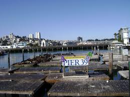
Ended up at the extremely busy and extremely touristy pier 39. It is kind of like a mix of a boardwalk and times square. Best part of the place had to be the sea lions that sit on docks next to the pier. There weren't too many, but that's because most of the sea lions go north to Alaska to the summer (I totally am a sea lion expert and didn't just read a sign).
3:50
Currently sitting again by the bay, this time with a great view of both Alcatraz and the Golden Gate Bridge. I find it strange how people were ok with a maximum security prison only a mile off the shores of a major city. I guess people just assumed that it was impossible to escape the island. I would have gone to visit Alcatraz, but a sign near the Alcatraz pier said that the next available tour was for August 16. I don't feel like waiting that long.

4:30
Made it over to Lombard street, AKA the crookedest street in America. It was a pain to get up here, as the hill is so steep that instead of walking up a road and being at a 45 degree angle, the city had to install stairs so it is easier to get up the hill. Once I got the the crooked part, I realized the street was less of a street and more of an amusement park ride, as there was a line of cars stretching for a few blocks, waiting to be able to drive down the street.

It's time for my visit to San Francisco to come to a close. I saw everything that I wanted to see, except for the Full House house, but it is too far away from anything else to get there. All in all, I really enjoyed my day in San Francisco.
12:29
First stop in San Fran is at this Burrito place in the Mission Neighborhood. It's supposed to have the best burritos in the city, and I would have to agree. It's amazing. Easily the best burrito I have ever had. Chopotle didn't have a chance.
1:36
Currently sitting under a tree in Union Square drinking a lemonade. It's weird how that right in the middle of the city there is this little enclave of nature. Might just chill here for a while it maybe go and shop a bit. There are a lot of stores surrounding the park, ranging from the extremely fancy ones with armed guards outside their doors to some that don't even have doors.
2:20
Finally made it over to the bay. Decided to take a little break, so I'm sitting on a dock on the bay, watching the tide roll away.
I was talking to my mom earlier and she told me that today in New Jersey it was 90+ with terrible humidity. That must suck, since right now it's 78 with nearly no humidity and a cool breeze. If there is one thing that I deffenitly don't miss from New Jersey it's the weather.

A really pretty panorama of the city.
3:00Ended up at the extremely busy and extremely touristy pier 39. It is kind of like a mix of a boardwalk and times square. Best part of the place had to be the sea lions that sit on docks next to the pier. There weren't too many, but that's because most of the sea lions go north to Alaska to the summer (I totally am a sea lion expert and didn't just read a sign).
3:50
Currently sitting again by the bay, this time with a great view of both Alcatraz and the Golden Gate Bridge. I find it strange how people were ok with a maximum security prison only a mile off the shores of a major city. I guess people just assumed that it was impossible to escape the island. I would have gone to visit Alcatraz, but a sign near the Alcatraz pier said that the next available tour was for August 16. I don't feel like waiting that long.

Look! It's the Golden Gate Bridge! It's so Gold(ish).
Made it over to Lombard street, AKA the crookedest street in America. It was a pain to get up here, as the hill is so steep that instead of walking up a road and being at a 45 degree angle, the city had to install stairs so it is easier to get up the hill. Once I got the the crooked part, I realized the street was less of a street and more of an amusement park ride, as there was a line of cars stretching for a few blocks, waiting to be able to drive down the street.

Look at how crooked it is! And the view of the city is really nice too.
5:30It's time for my visit to San Francisco to come to a close. I saw everything that I wanted to see, except for the Full House house, but it is too far away from anything else to get there. All in all, I really enjoyed my day in San Francisco.
Monday, July 20, 2015
Bridgid, Entry #4, The Doctor is (F)in
Hi everyone!
Hope that everyone’s summer in the lab is going well! This past week (the week of the 11th, sorry by last entry was a bit delayed) was my fourth week in the lab. I can’t believe that I am already more or about half way done my time with my lab, the days are flying by! This week was slightly more of a down week in the lab, running the standard procedures for new groups of embryos. (It was also a shorten week for me because we had a last minute family issue that I needed to care of, which required a shortened day on Wednesday and off on Thursday.)
This week on Monday, I started out the week with another day of imaging. This time, however, I imaged the 24 hpf embryos. It took a large part of the day, but I ended up recovering 20/25 embryos from the in situ for imaging. Since the 24hpf embryos are more developed and have more of a defined figure (aka look more like an actual fish), they were in my opinion, much easier and much more interesting to image than the 18 hpf. I honestly don’t know why, but personally I guess it made a different when they were more developed, maybe because they looked prettier (and more recognizable) in the pictures, but also because they were much easier to position. After I finished imaging in the afternoon, I started the first part of the DNA extraction for the other half (#9-19) of the 18 hpf embryos. Because the third step in the procedure for the DNA extraction is to incubate the embryos over night in the water bath, I wanted to make sure that I started today, so that I would have the DNA ready tomorrow.
On Tuesday morning, I came in and got right to work on the rest of the DNA extraction procedure for the last half of the 18hpf embryos. Once I had extracted the DNA, I measured the DNA on the Nanovue. Because I finished a little earlier than expected, I was able to go with Abby and Patrizia to the Influx machine at Penn. The Influx is very similar to the cell sorter (JACS) that we used last week, but it sorts the cells more gently, so that hopefully we will have cells on the slides after the cytosine and fixes the problem that we had last week. Having never used the Influx before, we just used a control group for the cell line, lmo2, that we were sorting, to make sure that it gave us cells at the end that were visible before we ran the actual experiment. After we took the cells sorted from the Influx to the cytospin, we were so happy to see that there were beautiful, clear cells on each of the slides!!! I spent part of the afternoon with Patrizia looking at the slides and the morphology of the cells. It was awesome to see so many different types of blood cell lineages at different stages of maturation under the microscope. I honestly could have spent hours looking and identifying all the cells, I thought it was that interesting. Also, it was so great to know that the Influx worked to sort the cells and gave us the results that Patrizia wanted! In addition to imaging the slides, I also ran the PCR for the samples that I did the DNA extraction for that morning so that they would be ready for sequencing this week.
Because of a family issue, I only came in Wednesday for a few hours before heading home. In that time, I ran a gel with my PCR from yesterday and then started to cut some of the bands from the gel for purification. Patrizia said that she would happily finish anything that I didn’t get to so that the PCR could be sent for sequencing. Unfortunately, when Patrizia went to finish cutting the gel, she noticed that the marker and some of the PCR products had faded and that the sequencing would most likely not go well. Since the results from the first 8 samples came back with an unreadable sequences anyway, we decided that it would just be better to re-run all the PCR for all 19 samples of the lmo2 18hpf DNA.
All day Friday was focused on re-running the PCR for all 19 samples of the lmo2 18hpf and then running the samples on a gel to prepare for sequencing. Also, because something might have been weird with the gel that I prepared (might have been too cold or hot when I added the ethidium bromide), Patrizia wanted me to watch her prepare the gel to see if I noticed anything different. On a different note, this is the first time that I have mentioned it, but because CHOP has so many summer students in labs across the campus, they have put together a really nice seminar series that features all types of doctors from all different departments at CHOP. Although most of the kids who attend the lectures are in college, I am lucky enough to attend the weekly sessions on Fridays with Abby. Each seminar runs about an hour long, with each doctor talking a little bit about their field and then talking a little bit about their journey that led to who they are now, along with giving career advice. The talks are always so informative and it is really cool to get a background in all different types cancer research that are going on at CHOP and the innovative techniques that so many of these doctors are creating and using to make huge strides in the cancer community. The career advice is also extremely valuable, especially for someone who is only in high school! That afternoon, I helped Abby with some imaging of a specific transcription factor, cmyb, in the MLL-GAS7 fish. Patrizia and I also prepared a gel to run my PCR, but unfortunately when I went to take out the gel from the model, it ripped. Because it was almost time for the lab meeting and we wouldn’t get the sequences until Tuesday regardless of if we ran the gel today or Monday, we decided that it would be better to not rush and finishing running the gel on Monday. Friday finished up with a lab meeting, where I got to learn more about the research that the other post docs in my lab are conducting and Abby and I also did a short presentation about our research this week!
Soo, Entry #4: Last Week
Since it is the last week of my internship in Dr. Bassett's lab, I am finishing up my works and re-organizing the results so that other people can later on easily understand my work.


I first went back to working with Gephi and generated brain network images with different modularity values. Small modularity value detects many small communities, whereas greater modularity value senses larger groups. The figures below are images of same subject and scan, but they have different modularity.
(modularity = 0.2)
(modularity = 1)
(modularity = 5)
After finalizing the gephi images, I revised my MatLab code so that I can save the values that I've used to generate the boxplots. I think the most important task that I would have to complete before I leave is to make sure that all results and data that I've worked with are saved and accessible.
Azza Entry #2 - The Other Side of the Lab
Last week I had the opportunity of shadowing my PI, Dr.
Rahul Deo, in the clinic to observe how he interacts with his patients, and the
type of concerns his patients bring to him. Yesterday we had a female patient
in her mid-sixties. She came to UCSF clinic seeking answers concerning her
cardiovascular state. Her older brother passed away a couple months after being
diagnoses with hypertrophic cardiomyopathy. Hypertrophic cardiomyopathy causes thickening
of the heart muscle linings making difficult for the heart to pump blood as
easily. In turn, making the heart significantly weaker. It turned out that most
of her family has died from heart failure and a majority of them being from
hypertrophic cardiomyopathy. Not knowing whether her relatives died from
inheriting a heart disease or simply due to age and higher blood pressure. Dr.
Deo has to gather genetic testings’ her relatives have done in Canada before
being able to tell whether this patient has also inherited a mutation for heart
failure. After meeting with the patient Dr. Deo showed me an ultra sound of the
patients’ hearts and pointed out to me the different chambers of the heart and
also showed me some thickening of the patient’s atrium, but he figured the
thickening could have been due to high blood pressure and age. He also showed me an EKG which is a electrocardiogram
of the patient and showed that there were no obvious reason that would validate
the patient of having a mutation. This was really interesting to me because not
only have I been able to work in the lab, but I also got to see how Dr. Deo
interacts with patients, and how patients like that influence future questions
and concerns in the world of cardiology.

Katie Entry #3: Finishing Growth Curves
Last week was my fourth week in the Cheung Lab. It was pretty similar to the previous week because I was finishing monitoring the growth of the potentially cold-sensitive mutant MRSA strains in optimal and cold conditions. I continued to observe the optical densities of each strain using a spectrophotometer, making growth curves using the ODs, and finding/comparing the growth rates of each strain at different temperatures . However, this week the postdoc that I have been working with, Dhana, found a better equation for determining the growth rate of cells using ODs so I went through my past and present work using that equation to find the doubling time of each culture and making new growth curves.
Also, some of the agar plates from the primary screening had issues growing in the incubator because of an issue with air flow on the top shelf so the agar on the plate ended up shrinking and becoming contaminated. I re-picked each strain on agar plates to grow at this temperature to finish the primary screening.
After I finished picking colonies and growing/observing the mutant strains in liquid media, I made a master graph to compare the growth of the 50 strains at each temperature and to pick the most important strains (the ones that grew the slowest in cold conditions) to use during transduction. We are going to start transduction next week using two strains so I went over the protocol and streaked the two strains that exhibited the most cold-sensitive phenotype on a plate to grow over the weekend.
Sunday, July 19, 2015
Emma, Blog #4, Weeks 5 and 6. WARNING GRAPHIC IMAGES
The past two weeks have really picked up in terms of what I have been doing in the lab.
Week 5 in the lab saw four big developments:
1. Finishing NeuN immunochemistry and moving to GFAP: NeuN immunochemistry is where you stain mice brains (at sizes of 50 micrometers) to view neuron channels under a microscope. Then, my mentor Yuling can run analyses on the brains in order to compare the amount of neurons between wild-type and genetically modified mice. I have been doing this type of immunochemistry for the past two weeks, but starting next week I am moving onto GFAP, which will stain brain slices to view glial filaments. The process between NeuN and GFAP immunochemistry is almost identical, however you use different secondary antibodies that bind to the primary.
2. Cutting brain slices: In order to start running GFAP, we need brain slices to stain. My mentor taught me how to use the machine that slices the brain slices to your desired thickness (which in our case is 50 micrometers). I spent about two hours cutting over 100 slices from one brain, which we then froze in a -80C freezer until we start the immuno next week.

3. Performing perfusion on three mice: Whole animal perfusion is a way to kill an animal and then fix it with paraformaldehyde to obtain the best preservation of the brain. This is an important process, because it needs to be done correctly in order to obtain brains that are preserved well and can be worked with either for staining or western blots. To perfuse a mouse, you first sedate it with ketamine threw a intramuscular injection. Once the mouse is fully under anesthesia, you pin it down stomach up in order to cut into the stomach. After cutting through the stomach you go up through the sternum to expose the heart. You inject a butterfly needle into the heart and pump in saline, and then quickly cut the heart to kill the animal. The saline is taken up through the capillaries and flushes out all of the blood. Once the animals lungs are white, and there is no longer any blood left in its system, you switch out the saline for paraformaldehyde, which is a fixative that preserves the brain instantly.
Here are some pictures from the procedure.


4. Joining a behavioral studies project: At lab meeting, my PI Dr. Siegel announced that he needed people to help in a behavioral study that tested Dextras1's effect on memory, EEG readings, and locomotion. I am specifically working on running 30 mice through a T Maze, a paradigm that tests working memory. I run the mice through 8 trials and then analyze the results.
Week 2
This week brought about another new project, as well as the start of my behavioral testing,
1. GFAP: This week I ran immuno for GFAP on brain's, and it went very well. My slices were all in tact with no breakage, which gave my PI the confidence to give me a project....
2. PROJECT: I got a project I am working on with one other high school student Meghan for the duration of my stay. I am using GFAP immuno staining to analyze if ketamine exposure in adolescent mice produces any differences in glial filaments when compared to wild mice. This may not seem related to schizophrenia, the labs main focus, however schizophrenia is a NMDA antagonist and is thought to have a correlation to the disease. I will start immuno next week for this project.
3. Behavioral testing: I ran my first trials on all 30 mice this week in the T maze. It took me the full day, but it was actually pretty fun. This was the first time I got to interact with the mice completely alone, which made me nervous at first but in the end it was fine. Next week I will have two more testing days before I can start analysis.
3. Clinical trials with my PI: Dr. Siegel not only runs a lab, but he meets with patients every Wednesday afternoon at UPenn's hospital. I was able to shadow him for the whole afternoon. Unfortunately, I cannot divulge much information because of HIPAA, but it was a great experience and I got to actually meet people dealing with schizophrenia. It was a really moving experience and made me glad that I was working in a lab that was trying to help people like them live a normal life again.
Jay Ha #2 growing e coli and collecting data
This week had been full of bacteria.
Now that I am sitting in front of my laptop trying to get my weekly journal stuff done, I wish I had taken more photos. I guess I will take more photos next week.

This is one of the few photos I took before autoclaving these bacteria and saying goodbye to my weekly production of lovely bacteria . As you can see, there are many plates, and all of these plates contain somewhere between 0 to tens of thousands of colonies of E Coli bacteria that I had grown. YaY!
My lab works started with production of more phosphate buffer solution and LB Broth. It wasnt that hard as I had to put some pills or powdery materials into the beaker using precise scientific scales and mixing them with DI water and shake them and put them in sonic machine that evenly mixes the solution. All solutions prepared had to be autoclaved to create a sterile environment. The phosphate buffer solution would be used in serial dilution method of E Coli culture to prepare E Coli of varying concentration. Some of the LB Broth, or the source of food for bacteria, are used to directly culture E Coli population in a shaking incubator set to 37.5 degrees Celcius, and other portions of LB Broth are mixed with solidifying agent so that they would be prepared to plates as seen in the picture above for the grown E Coli cultures to be tested for their growth pattern.
Plates were prepared under bio safe fume hood. Making forty plates is tedious but does not take that long time. When the E Coli culture that used to be frozen is defrozen and incubated and all prepared, they are serially diluted to the factors of 1, 10^-2, 10^-4, ....., 10^-8. This is done by adding 0.1 mL of E Coli culture or serially diluted cultures into 9.9 mL of phosphate buffer.
Then, these serially diluted solutions containing E Coli are spread onto the plates using glassware that spreads the culture onto the plates. Five sets of plates are prepared on every two hour intervals for 20 hours and put in the incubator to be counted in next 24 hours. After 24 hour intervals these plates are counted for the number of colonies of bacteria that had grown overnight.
OD measurements using spectroscopy machine are taken in varying time intervals to see the correlation between OD measurements and CFU.
I have prepared and counted the bacteria for five sets of two hours intervals everyday and had conducted this for four days total.
First sets of data were discarded because bacteria were poorly cultured because the incubator was partially broken, so the data taken from third and fourth days are below.
|
Time (h) |
OD at 600nm |
Count |
Dilution Factor |
CFU/mL |
|
0
|
-0.0048
|
335
|
1
|
3.35E+03
|
|
2
|
-0.0018
|
301
|
1
|
3.01E+03
|
|
4
|
-0.0063
|
512
|
1
|
5.12E+03
|
|
6
|
-0.0223
|
45
|
100
|
45000
|
|
8
|
-0.0021
|
309
|
100
|
309000
|
|
10
|
0.0123
|
61
|
10000
|
6.10E+06
|
|
12
|
0.771
|
165
|
1000000
|
1650000000
|
|
13
|
0.8125
|
|
|
|
|
14
|
0.4506
|
290
|
1000000
|
2900000000
|
|
15
|
0.8439
|
|
|
|
|
16
|
0.9864
|
161
|
1000000
|
1610000000
|
|
17
|
1.0974
|
|
|
|
|
18
|
1.1149
|
301
|
1000000
|
3010000000
|
|
19
|
1.196
|
|
|
|
|
20
|
1.208
|
261
|
1000000
|
2610000000
|
Here are three graphs from data collected this week.

Next week, I will be conducting research that deals with disinfection that had been catalysed through oxidation processes. Sounds fun. lol
Subscribe to:
Posts (Atom)